This repository contains some test scripts which use the NeuroPAL datasets.
Note: Work in progress! Subject to change without notice! Please get in contact if you would like to reuse this dataset, to help improve the quality of the conversion
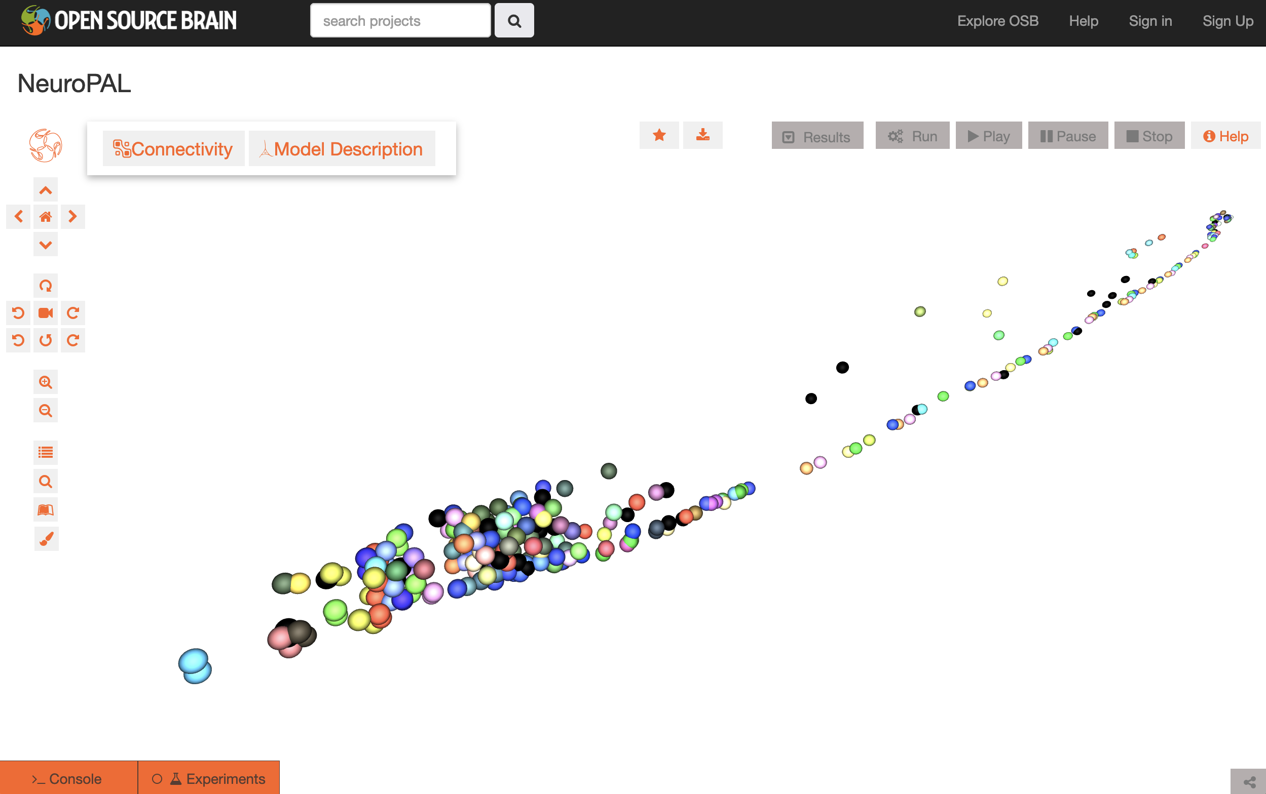
A canonical set of positions for the cell bodes as well as colors reflecting expression levels has been produced. A description of the data can be found in: Toward a more accurate 3D atlas of C. elegans neurons, Michael Skuhersky, Tailin Wu, Eviatar Yemini, Amin Nejatbakhsh, Edward Boyden & Max Tegmark BMC Bioinformatics volume 23, Article number: 195 (2022)
More details on the data can be found here, and a Jupyter notebook containing the conversion of this data to NeuroML is here.
The above image shows the canonical positions and colors visualised on Open Source Brain.
The original publication on NeuroPAL was: Eviatar Yemini, Albert Lin, Amin Nejatbakhsh, Erdem Varol, Ruoxi Sun, Gonzalo E. Mena, Aravinthan D.T. Samuel, Liam Paninski, Vivek Venkatachalam, Oliver Hobert, NeuroPAL: A Multicolor Atlas for Whole-Brain Neuronal Identification in C. elegans, Cell, Volume 184, Issue 1, 2021.
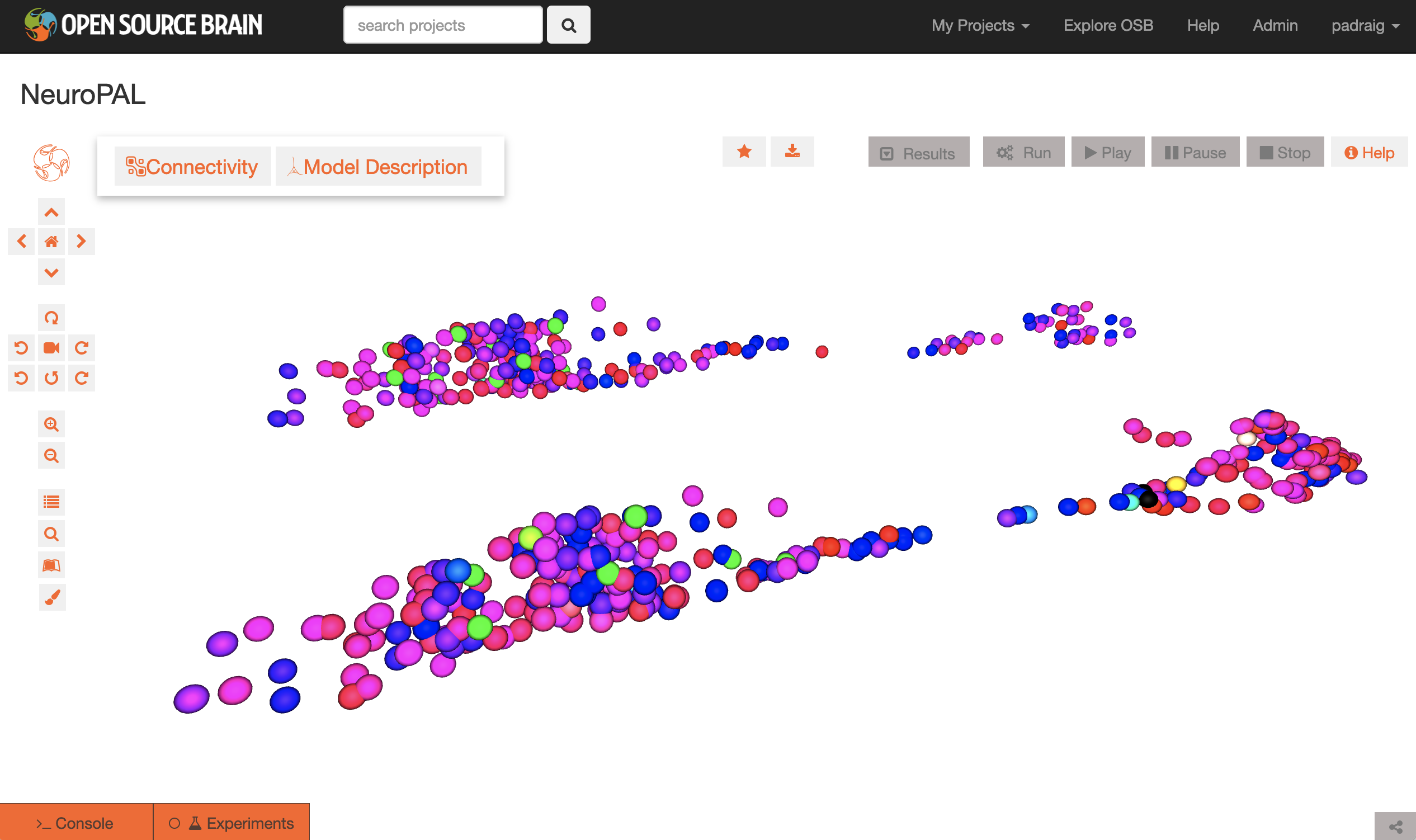
The dataset released with this publication on positions and colors (as used in Figures 2 and 7) of cells in the head and tail has been used in this Jupyter notebook, and extracted from the Excel sheet, and converted to NeuroML format.
The above image shows the data visualised on Open Source Brain, with the data from the male worm shown in front (head to left, tail to right, shifted to be adjacent), and the equivalent hermaphrodite data behind.
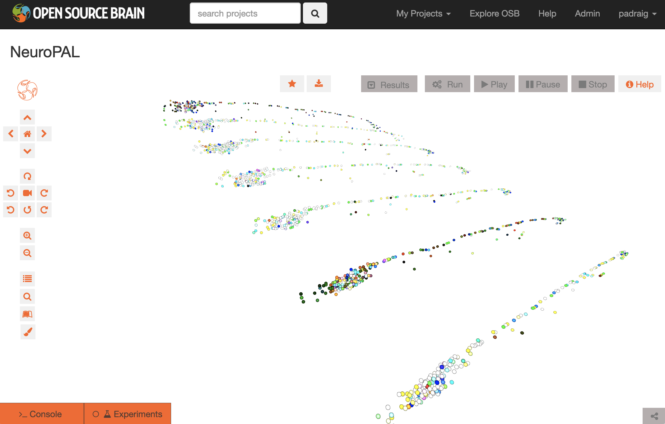
Updated data has been provided by Michael Skuhersky on the whole body cell positions and colors. Conversion of this data can be seen here.
The above image shows the whole worm data visualised on Open Source Brain. Seven worm reconstructions are shown, and the worm body/layout has been straightened/standardized as described here.


